Maximum quantity allowed is 999
Please select the quantity

ELISA Using Biotinylated Glycans
Enzyme-linked immunosorbent assay (ELISA) is a highly sensitive quantitative method for detecting specific molecules using antigen-antibody reactions. ELISAs are used in a wide range of fields including immunology, biomarker discovery and quality control. ELISAs can also be used to detect a wide range of molecules. The following are our ELISA protocols using anti-glyco antibodies, HSA glycan conjugates and biotinylated glycans.
Reagents and Materials
- Streptavidin from Streptomyces avidinii (Product No. S0951)
- Biotinylated glycans: L2-L2-β-PEG3-biotin (Product No. G0516), L4-L4-β-PEG3-biotin (Product No. G0517)
- Coating buffer: 0.1M Carbonate Buffer (pH8.5)
- Blocking solution: 1% BSA/TBST
- Plate washing buffer: TBST (Tris-buffered saline with 0.05% Tween20)
- HRP Chromogenic substrate solution: TMB solution (Product No. T3854), etc
- Primary antibodies: Anti-6-sulfo LacNAc Monoclonal Antibody (AG105) (Product No. A3251), Anti-6,6'-disulfo LacNAc Monoclonal Antibody (L4L4-8) (Product No. A3252)
- Secondary antibody: Goat Anti-Mouse IgG+M Secondary Antibody
Procedure
- Add 100 μL of streptavidin solution (prepared at 10 μg/mL) to a high-binding ELISA plate and incubate for 2 hours at room temperature or overnight at 4°C.
- After washing the wells, add 150 μL of blocking solution to each well. Incubate for 2 hours at room temperature or overnight at 4°C.
- After washing the wells, add 50 μL of biotinylated glycan diluted to 0.2 μM with blocking solution to each well. Incubate for 1 hour at room temperature.
- After washing the wells, add primary antibody diluted with blocking solution to each well. Incubate for 1 hour at room temperature.
- After washing the wells, add secondary antibody diluted 5,000-fold with blocking solution to each well. Incubate for 30 minutes at room temperature.
- After washing the wells, add 100 μL of HRP chromogenic substrate solution and allow to react for 30 minutes.
- Add 100 μL of reaction stop solution and measure absorbance at 450 nm (Ref. 630 nm) with a plate reader.
Experimental Example
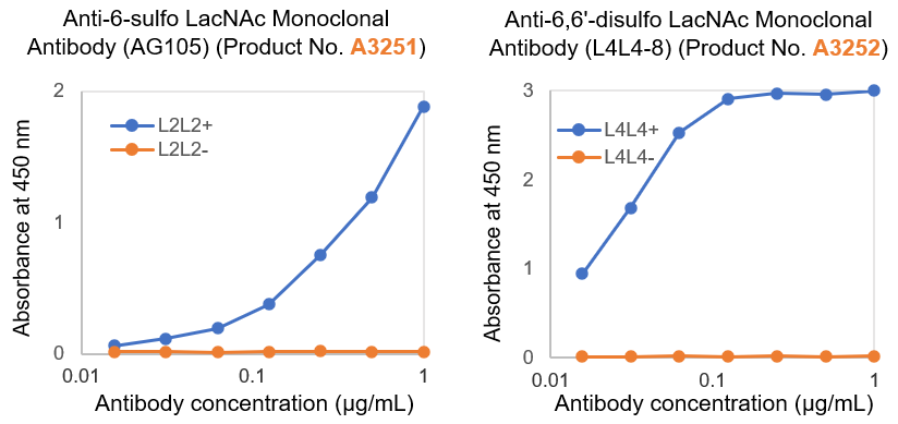
Antibody binding analysis by using biotinylated glycans
Related Products
ELISA Using HSA-Glycoconjugate
Reagents and Materials
- Human Serum Albumin-Gb5 (= HSA-Gb5) (Product No. H1777)
- HSA (Human Serum Albumin)
- Coating buffer: 0.1 M Carbonate Buffer (pH 8.5)
- Blocking solution: 1% BSA/TBST
- Plate washing buffer: TBST (Tris-buffered saline with 0.05% Tween 20)
- HRP Chromogenic substrate solution: TMB solution (Product No. T3854), etc.
- Primary antibodies: Anti-SSEA-3 (Gb5) Monoclonal Antibody (Product No. A3729)
- Secondary antibody: Goat Anti-Mouse IgG+M HRP Conjugate

Chemical Structure of HSA-Gb5 (Product No. H1777)
Procedure
- Add 100 μL of HSA-Gb5 solution (prepared at 1 μg/mL) to a high-binding ELISA plate and incubate for 2 hours at room temperature or overnight at 4°C.
- After washing the wells, add 150 μL of blocking solution to each well. Incubate for 2 hours at room temperature or overnight at 4°C.
- After washing the wells, add primary antibody diluted with blocking solution to each well. Incubate for 1 hour at room temperature.
- After washing the wells, add secondary antibody diluted 5,000-fold with blocking solution to each well. Incubate for 30 minutes at room temperature.
- After washing the wells, add 100 μL of HRP chromogenic substrate solution and allow to react for 30 minutes.
- Add 100 μL of reaction stop solution and measure absorbance at 450 nm (Ref. 630 nm)with a plate reader.
Experimental Example

Specific detection of Gb5 by using anti-SSEA-3 (Gb5) antibody
Related Products
Glycosphingolipid ELISA
Reagents and Materials
- Glycolipid solution (Dissolved at 1 μg/mL in ethanol)
- Blocking solution: 1% BSA (bovine serum albumin) in PBS
- Plate washing buffer: PBS (phosphate-buffered saline)
- HRP Chromogenic substrate solution: TMB solution (Product No. T3854), etc.
- Primary antibodies: Anti-Globo-H Monoclonal Antibody (Product No. A3703), Anti-SSEA-4 (SialylGb5) Monoclonal Antibody (Product No. A3742), Anti-SSEA-3 (Gb5) Monoclonal Antibody (Product No. A3252), Anti-Gb4 Monoclonal Antibody (Product No. A3776), Anti-Gb3 Monoclonal Antibody (Product No. A2506)
- Secondary antibody: Goat Anti-Mouse IgG+M HRP Conjugate
Procedure
- Add 100 μL of glycolipid solution (1 μg/mL) to an ELISA plate and dry completely at 37°C (for 3 hours to overnight).
- Add 300 μL of blocking solution to each well and incubate for 2 hours at room temperature or overnight at 4°C.
- After washing the wells, add primary antibody diluted with blocking solution. Incubate for 30 minutes at room temperature.
- After washing the wells, add secondary antibody (diluted 1:10,000 with blocking solution). Incubate for 30 minutes at room temperature.
- After washing the wells, add 100 μL of HRP chromogenic substrate solution and allow to react for 30 minutes.
- Add 100 μL of reaction stop solution and measure absorbance at 450 nm (Ref. 630 nm) with a plate reader .
Experimental Example

Specificity analysis of antibodies using various glycolipids
Related Products
Detection of Chondroitin Sulfate A by Flow Cytometry
Glycosaminoglycans (GAGs) are polysaccharides that comprise repeated disaccharide structures of amino sugar and either uronic acid or galactose found on the surface of cells and in the extracellular matrix. Chondroitin sulfates are one example of GAGs; they can be found in various tissues such as cartilage, corneal tissue, and the brain, and are involved in various phenomena including moisture regulation, cell adhesion, and nerve regeneration. Roles for chondroitin in diseases such as breast cancer have also been the object of studies. As the instrumental analysis of polysaccharides, including GAGs, is known to be difficult, specific antibodies against GAGs are extremely useful as a detection tool. Flow cytometry (FCM) makes the expression analysis of target antigens such as GAGs in target cells quick and easy through the use of fluorescence-labeled antibodies, allowing for the characterization of individual cells.
Reagents and Materials
- MCF-7 cells (* MCF-7 cell line (JCRB0134, H. D. Soule et.al.) was obtained from JCRB Cell Bank.)
- Mouse IgM isotype control (10 μg/mL dilution)
- Anti-Chondroitin Sulfate A Monoclonal Antibody (LY111) (Product No. A3143) (10 μg/mL dilution)
- Goat Anti-Mouse IgM R-PE Conjugate (2.5 μg/mL dilution)
- Phosphate-Buffered Saline (PBS)
- Trypsin/EDTA
- Cell culture medium for MCF-7
- 0.5% BSA-containing PBS
Procedure
- Culture MCF-7 cells.
- On the day of the experiment, wash cells with PBS, trypsinize them, and neutralize with complete culture medium.
- Wash cells twice with 0.5% BSA-containing PBS.
- Resuspend cells in 0.5% BSA-containing PBS and divide into two tubes so that each tube contains 2x10⁵ cells (100 μL).
- Add either mouse IgM isotype control or anti-chondroitin sulfate A monoclonal antibody, mix, and incubate for 30 minutes at 4°C.
- Wash cells three times with 0.5% BSA-containing PBS.
- Resuspend cells in 100 μL of goat anti-mouse IgM R-PE conjugate diluted with 0.5% BSA-containing PBS, and incubate for 30 minutes at 4°C under the dark.
- Wash cells three times with 0.5% BSA-containing PBS, and analyze via flow cytometry.
Experimental Example

Flow Cytometric Analysis.
MCF-7 cells were reacted with anti-Chondroitin Sulfate A antibody (Product No. A3143) (red line) or isotype control (blue line),
then stained with fluorescent secondary antibody and analyzed by flow cytometer.