

Published TCIMAIL newest issue No.199
Maximum quantity allowed is 999
Bitte wählen Sie die Menge aus.
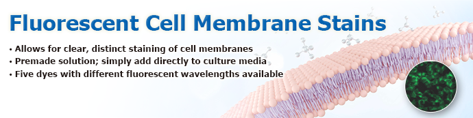
The cell membrane defines the boundary between a cell's interior and its surroundings, playing key roles in signal transmission and molecular transport. We offer five membrane-selective fluorescent dyes that enable clear, consistent staining with minimal effort; simply add the reagent to your culture media. Each dye emits in a unique wavelength range, allowing for multiplexing with other fluorescent probes.
Products
Maximum Excitation/Emission Wavelength of Fluorescent Stains
| Fluorescent Stains | Maximum Excitation Wavelength (nm) | Maximum Emission Wavelength (nm) |
|---|---|---|
| DiI Solution (Product No. D6202) | 553 | 575 |
| DiD Solution (Product No. D6263) | 657 | 678 |
| DiO Solution (Product No. D6539) | 491 | 509 |
| DiR Solution (Product No. D6540) | 757 | 781 |
| PKH 26 Solution (Product No. P3316) | 554 | 573 |
Example of Membrane Staining with DiI Solution (Product No. D6202)
- Culture cells and remove medium.
- Add medium supplemented with D6202 to a final concentration of 5 μM (staining medium), and incubate cells at 37°C for 10 minutes.
- Remove staining medium and wash three times with PBS(-).
- Observe cells via a fluorescence microscope.
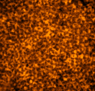
Figure 1. HeLa cells stained with D6202
Example of Membrane Staining with PKH 26 Solution (Product No. P3316)
- Culture cells and remove medium.
- Add medium supplemented with P3316 to a final concentration of 2 μM, and incubate cells at room temperature for 5 minutes.
- Add a volume of fetal bovine serum (FBS) equivalent to the volume of medium used in step 2, and incubate cells at room temperature for 1 minute.
- Remove medium with FBS and wash three times with PBS(-).
- Observe cells via a fluorescence microscope.
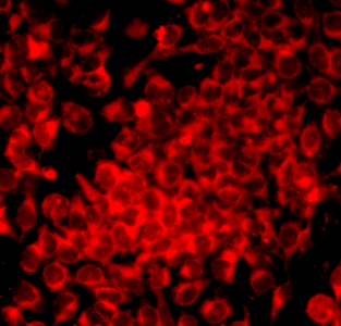
Figure 2. HeLa cells stained with P3316