

Maintenance Notice (4:00 AM November 1 - 9:30 AM November 1, 2025): This website is scheduled to be unavailable due to maintenance. We appreciate your patience and understanding.
Published TCIMAIL newest issue No.199
Maximum quantity allowed is 999

Cancer is a disease caused by accumulation of genetic mutations and epigenetic alterations, leading to abnormal cell proliferation and subsequent tumor cell escape from immune surveillance. In highly malignant tumors, cancer cells invade surrounding tissues and metastasize to distant organs. While death can be prevented by early diagnosis and treatment in some cancers, other cancers continue to show poor prognoses. It is therefore critically important to develop and establish better diagnostic methods and treatments for such cancers.
Glycans can be found on the surface of cells and within tissues, and play essential roles in cellular attachment, cell recognition, intra- and inter-cellular signaling, and embryonic development. It has been found that structures and expression level of various glycans are altered in cancer cells and cancer tissues. As these changes have been linked to disease progression, these glycans hold promise as diagnostic markers and therapeutic targets.
We offer a wide variety of glycan-related reagents useful for cancer research.
‡All the products in this page are reagents and for research use only.
Cellular glycosylation profiles significantly change during carcinogenesis. Such altered cellular carbohydrate structures are known as tumor-associated carbohydrate antigens, and some are used as tumor markers. Tumor-associated carbohydrate antigens are considered promising targets in immunotherapy as well as in vaccines and therapeutic antibody development.
We support the research in this field with various tools.
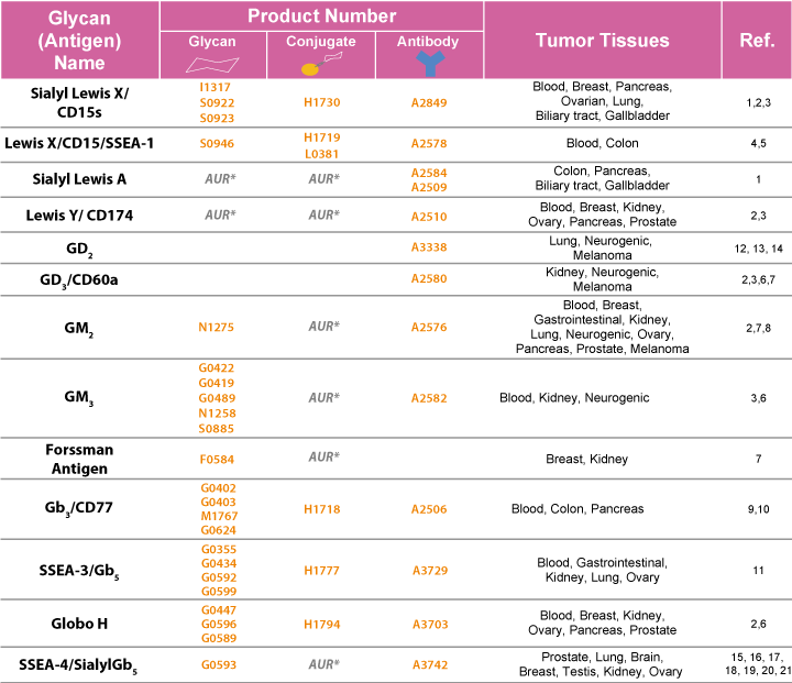
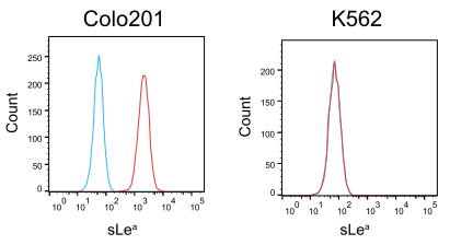
Sialyl Lewis A (sLea) is a carbohydrate antigen also known as a tumor marker CA19-9.
Colo201 and K562 cell lines were stained with either Anti-Sialyl Lewis A Monoclonal Antibody (1H4) (Product No. A2584) (red line) or an isotype control (blue line). Then, the cells were stained with Goat Anti-Mouse IgG R-PE Conjugate (Product No. G0569), and finally analyzed by flow cytometry. sLea was detectable in Colo201 but not in K562.
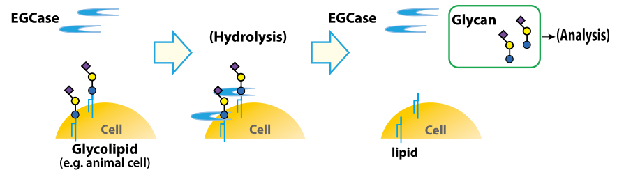
The ability to analyze alterations of cellular glycosylation profile is highly important for a complete understanding of cancer. We have an extensive line-up of various reagents useful in the analysis of carbohydrates, such as lectins to detect glycans, endoglycosidases to cleave oligosaccharides, and chemically synthesized oligosaccharides.
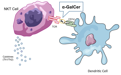
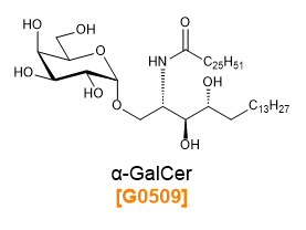
α-Galactosyl Ceramide (α-GalCer) is an artificial glycolipid developed based on a glycosphingolipid originally extracted from the sea sponge, Agelas mauritianus.22-24) α-GalCer forms a complex with antigen presenting molecule CD1d and serves as a ligand for TCR on NKT cells (cells with both NK cell- and T cell-like characteristics). This complex activate NKT cells and cause them to produce various cytokines, which in turn give rise to both immune-activating and immune-suppressive responses.25-28) Because of this, α-GalCer has been used in cancer immunotherapy research.29)
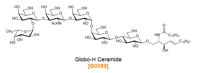
In addition to α-GalCer, we also offer other chemically synthesized oligosaccharide antigens useful for cancer vaccine and other immunotherapy-based research fields.
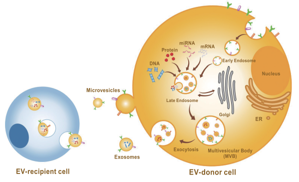
Extracellular Vesicles (EVs) are small vesicles composed of a lipid bilayer which are secreted from various cells. Produced under both physiological and pathological conditions, EVs can be found in bodily fluids as well as cell culture supernatants, where they are taken up into target cells. Packaged within EVs can be found functional molecules such as nucleic acids and proteins. In addition, EVs are known to present surface glycans, and are deeply involved in cancer metastasis.
As cancer metastasis is a major contributor to poor prognoses, its control is of critical importance for successful cancer therapy. The extracellular matrix plays important roles in tumor invasion and metastasis. Hyaluronic acid, its major polysaccharide component, acts via its receptor CD44 to activate intracellular signaling molecules such as those related to cell migration, leading to cancer progression.30)