

Maintenance Notice (5:30 - 10:30 PM June 28, 2025): This website is scheduled to be unavailable due to maintenance. We appreciate your patience and understanding
Published TCIMAIL newest issue No.198
Maximum quantity allowed is 999


(Laser Scanning Microscope: Olympus FLUOVIEW FV 3000)
Green: α-Tubulin,Blue: Nuclear DNA
The HeLa cells were fixed with 4% paraformaldehyde, and permeabilized with 0.1% Triton X-100.
Then, the specimen was blocked in BSA/PBS blocking buffer.
Subsequently, the specimen was incubated with properly diluted primary antibody (Mouse Anti α-Tubulin IgG). And, after washing, was further incubated with biotin-conjugated secondary antibody (Goat Anti-Mouse IgG Biotin Conjugate [G0387]).
Finally, it was rinsed with PBS and stained with each fluorescent-probe*.
*Streptavidin FITC Conjugate [S0966] at 5 µg/mL (shown in green), DAPI·2HCl [A2412] at 1 µg/mL (shown in blue)

(Laser Scanning Microscope: Olympus FLUOVIEW FV 3000)
Red: α-Tubulin, Green: Endoplasmic Reticulum, Blue: Nuclear DNA
The HeLa cells were fixed with 4% paraformaldehyde, and permeabilized with 0.1% Triton X-100. Then, the specimen was blocked in BSA/PBS blocking buffer.
Subsequently, the specimen was incubated with properly diluted primary antibody (Mouse Anti α-Tubulin IgG and Rabbit Anti KDEL** IgG).
Finally, it was rinsed with PBS and stained with each fluorescent-probe***.
** KDEL (amino acid sequence) normally localize in the endoplasmic reticulum.
*** Goat Anti-Mouse IgG R-PE Conjugate [G0569] at 5 µg/mL (shown in red),
Goat Anti-Rabbit IgG FITC Conjugate [G0452] at 5 µg/mL (shown in green),
Hoechst 33258 [H1343] at 1 µg/mL (shown in blue)

(Flow cytometer: Sysmex RF-500)
The HeLa cells were incubated with Mouse Anti CD9 Antibody (red line) or Mouse IgG2aκ isotype control (black line) at 5 µg/mL.
Subsequently, both were stained with Goat Anti-Mouse IgG FITC Conjugate [G0406] at 5 µg/mL.
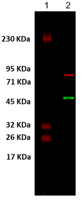
(Imager: Cytiva Amersham Imager 680)
Red: Calnexin, Green: α-Tubulin
Lane 1: Color Protein Marker, Lane 2: HeLa Whole Cell Lysate at 5 µg
Transfer of HeLa cell lysate to PVDF membrane. Then, the specimen was blocked in BSA/TBST blocking buffer.
Subsequently, the specimen was incubated with properly diluted primary antibody (Mouse Anti Calnexin IgG and Rabbit Anti α-Tubulin ).
Finally, it was rinsed with TBST and stained with each fluorescent-probe*.
*Goat Anti-Rabbit IgG FITC Conjugate [G0452] at 5 µg/mL(shown in green)
Goat Anti-Mouse IgG R-PE Conjugate [G0569] at 2 µg/mL (shown in red)


Acridine orange is a nucleic acid staining dye that is used to identify dead cells. It intercalates with the double-stranded DNA base pairs at a ratio of 1:3 and is capable of emitting green fluorescence (Ex: 500 nm, Em: 520 nm). It also emits red fluorescence (Ex: 460 nm, Em: 650 nm) when bound to RNA or single-stranded DNA. Since acridine orange is mutagenic, this product is supplied as a solution that prevents it from splashing during weighing.

NIH3T3 cells stained by A3396
Please adjust the staining duration and the volume of the solution according to the cell density.
Some cells may require prior fixation; therefor optimization of the protocol according to your need is recommended.

λDNA was stained using either the acridine orange solution [A3396] or the other manufacturer's product, and the fluorescence measured from two experiments was compared (Ex:500nm, Em:520nm).
The results indicated that A3396 stained better than the other manufacturer's product.

Figure. Fluorescence in mitochondria in mouse peritoneal neutrophils. (Provided by Prof. Kobayashi)
S. Sasaki, S. Yamada, M. Iwamura, Y. Kobayashi, Free Radic. Biol. Med. 2013, 65, 1005.

Excitation wavelength λex, max = 335 nm
Emission wavelength λem, max = 616 nm
Sharpened emission spectrum
Large Stokes shift (the difference in wavelength between positions of the band maxima of the absorption and emission spectra)

Available in Tris, TE, PBS, etc. for wide use
Time-resolved fluorometric measurement can remove background fluorescence from the sample matrix and often gives detectability better than one order of magnitude compared to those of conventional fluorometric assays.
Note: Sensitive detection of DTBTA-Eu3+ labeled probes requires time-resolved fluorometric measurements.
